Non-surgical TMJ treatment and functional assessment
I assess and treat problems affecting the jaw joints and chewing muscles.These include jaw pain, painful clicking, restricted mouth opening and symptoms associated with bruxism.
TMJ problems may involve pain, altered jaw movement or muscle tension. Tell me when your symptoms started, how they change during the day and whether they affect eating, speaking or sleep.
Clicking, grating and locking
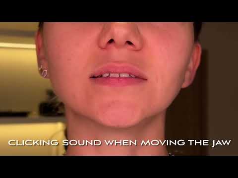
A joint sound needs to be considered alongside pain and movement. Does it happen every time? Does your jaw catch? Is eating becoming difficult? Painless clicking with comfortable, unrestricted movement usually does not need treatment. Pain, repeated locking or increasing difficulty opening your mouth are reasons to have an assessment.
Clicking during jaw movement
Difficulty opening your mouth or a jaw that shifts to one side
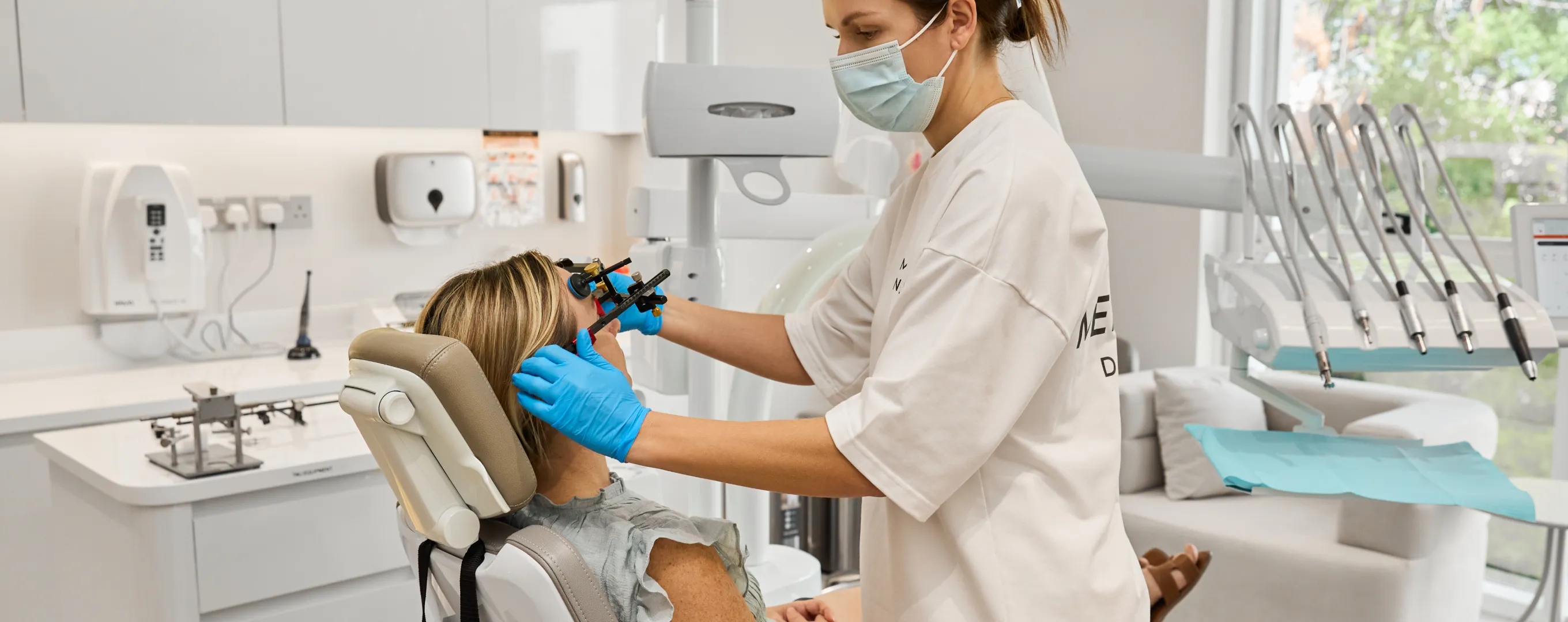
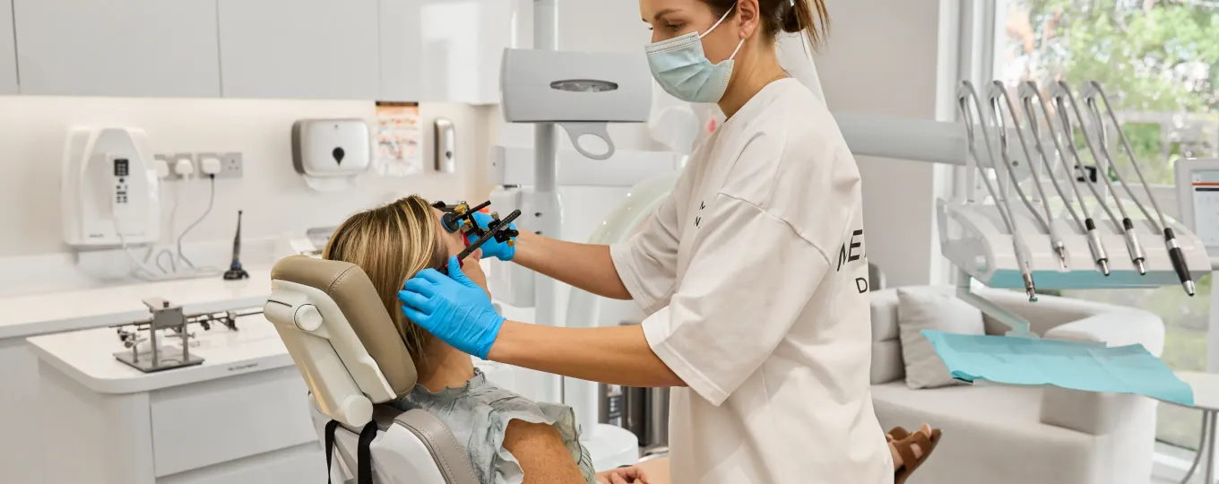
Tell me if your mouth opens less than it used to, your jaw moves noticeably to one side or you need to find a particular position to open wider. I assess the range and symmetry of movement, tenderness and the point at which movement becomes difficult. In selected cases, an instrumental recording can help examine the movement in more detail.
Restricted mouth opening
Jaw pain, temple pain and aching muscles
Pain may be felt in front of the ear, in the cheek or around the temple. It may become worse when chewing, talking for a long time or opening wide. Some people mainly notice morning stiffness: the jaw feels tired or tense before breakfast. During the examination, I check which movements and areas of tenderness reproduce the pain you recognise.
Bruxism, worn teeth and chipped restorations
Night-time grinding, daytime clenching, tired jaw muscles and repeated chips in dental work are reasons to assess the load on your teeth. I examine teeth, fillings and crowns and ask about clenching and how your jaw feels after sleep. Tooth wear may have developed over a long period, so I compare it with your current symptoms rather than treating it as proof of active bruxism.
Tinnitus (ringing in the ears)
Tinnitus is a ringing, buzzing or other sound heard without an external source. It can accompany TMJ disorders, but it also has many other possible causes. If the sound changes when you move your jaw or clench your teeth, mention this at your consultation. Identifying the cause involves assessing jaw function alongside a hearing assessment and ENT consultation.
Ear symptoms, facial discomfort and neck tension
Some patients also report pain near the ear, fullness, headaches or neck tension. Mention these symptoms, particularly if they change when you move your jaw. They can have other causes, and a TMJ assessment should complement any necessary ENT, neurological or other medical assessment.
An injury, significant swelling, inability to eat or drink, or sudden severe difficulty moving the jaw needs prompt in-person care.
Causes
What is TMJ, and what causes TMJ disorders?
The temporomandibular joint (TMJ) connects the lower jaw to the temporal bone of the skull. You have two of these joints, one just in front of each ear. Together with the chewing muscles, they allow you to open and close your mouth, chew and speak.
A disc sits between the joint surfaces, helping distribute load and allowing the joint to move smoothly.
Temporomandibular disorders (TMD) is the collective name for conditions affecting these joints and the chewing muscles. They can cause jaw pain and difficulty moving the jaw. Their causes and mechanisms vary; the examples below explore bruxism and joint instability.
Bruxism and strain on the jaw
Clenching and grinding can leave the chewing muscles tired or sore and can damage teeth and restorations. I ask when it happens, how your jaw feels after sleep and whether you notice daytime clenching. These details help relate the load on your jaw to your symptoms.
The animation illustrates grinding and changes within the joint. It does not mean that everyone who grinds their teeth develops TMD or a displaced disc. Those findings need to be assessed individually.
Bruxism and TMD: a 3D animation of teeth grinding and disc displacement.
Jaw instability and dislocation
Some people experience episodes in which the jaw slips out of position or gets stuck after opening wide. A dislocation involves the joint head moving out of its normal working position; it is different from displacement of the disc inside the joint. Clicking alone does not establish that a dislocation has occurred.
I ask about previous episodes, injuries and difficulty closing the mouth. The animation explains the mechanics of dislocation. If your mouth is stuck open and you cannot close it, seek urgent in-person care rather than waiting for a routine appointment.
Jaw dislocation: a 3D animation of joint instability.
Other factors and overlapping symptoms
For one patient, muscle pain and clenching may be prominent. Another may have restricted movement, changes within the joint or a history of injury. Sleep, stress and persistent pain can also affect symptoms. Sometimes pain comes from a tooth or nearby tissue but feels as though it is in the joint.
I also look for tooth wear, missing teeth, damaged restorations and changes in how the teeth meet. This helps identify dental problems that need care alongside the joints and muscles.
If you have jaw pain, clicking or difficulty opening your mouth, you may search for a “TMJ specialist in Dubai”. You are looking for a clinician experienced in jaw joint and chewing muscle problems. Their licensed specialty, further training and clinical experience help you understand whether their practice fits your needs.
I am a Specialist Prosthodontist, with a clinical focus on functional assessment and conservative care for TMJ disorders. My background includes research into TMJ diagnosis and further training at VieSID.
Why see a prosthodontist for TMJ problems?
DHA includes the diagnosis and management of TMJ dysfunction within a prosthodontist’s scope of practice. I assess the joints, chewing muscles and how the teeth meet, then recommend care based on the findings. This may include splint therapy and input from other clinicians when needed.
When is an oral and maxillofacial surgeon needed?
If the joint condition needs a surgical assessment, I refer the patient to an oral and maxillofacial surgeon. The surgeon assesses whether a procedure is appropriate and performs it with the necessary privileges. The recommendation may also be to continue conservative care.
After splint therapy, we review your progress and the condition of your teeth. If damaged or missing teeth need restoring, or orthodontic treatment is needed, I explain why and discuss the benefits, risks and alternatives.
The assessment starts with a conversation and a diagnostic questionnaire. I ask when the symptoms started, where you feel pain, whether your jaw clicks or locks and which movements are difficult. Previous injuries, dental treatment and investigations help establish what has changed.
The clinical examination includes opening and sideways movements, the jaw joints and chewing muscles, the teeth and how they meet. Photographs and digital models, when needed, document the starting point for planning.
After the examination, I explain which further information we need about bone, the disc and soft tissues, or jaw movement. The sections below describe how we obtain it.
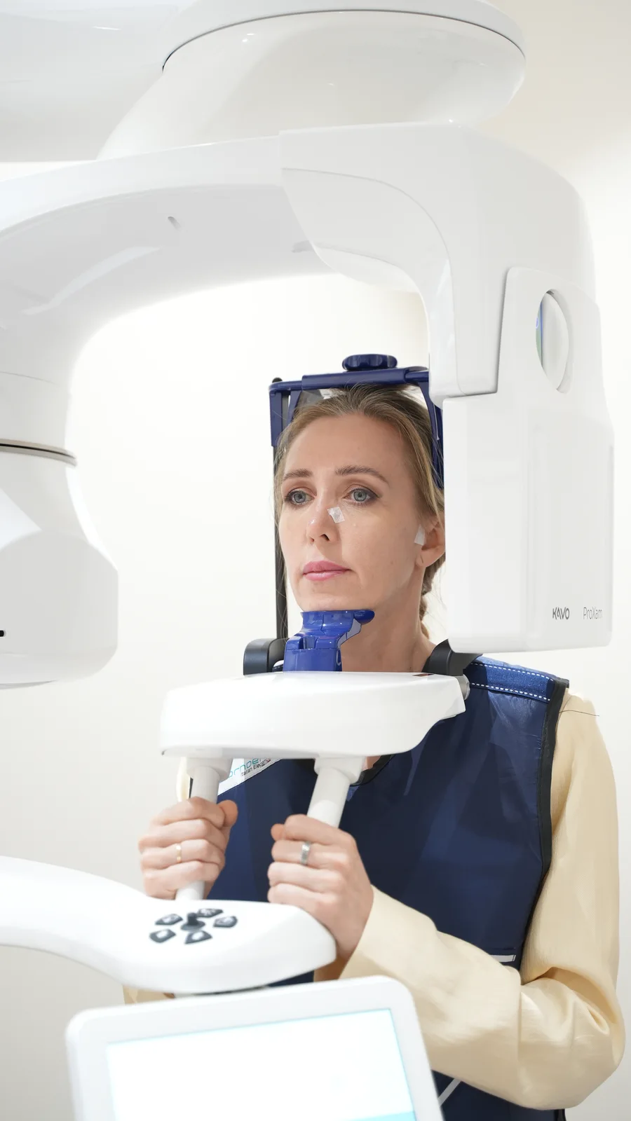
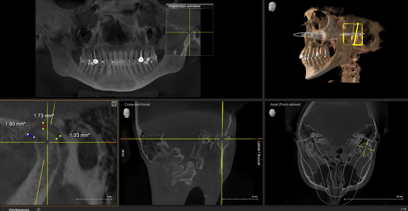
Cone-beam computed tomography (CBCT) or computed tomography (CT) provides important information about the bony structures of the joint. These scans help assess joint surface shape and bone changes. I decide which imaging is needed after the examination, taking existing scans into account.
A conversation, diagnostic questionnaire and examination first. Further investigations when indicated.
Imaging addresses specific clinical questions; it is not needed in every case.
CBCT of the temporomandibular joint. Different views help assess the bony structures and their relationship. The enlarged section shows an example of joint-space measurements. Select the image to view it in detail.
Clinical screening
Hamburg screening: what the clinician checks
The Hamburg screening (CMD-Kurzbefund by Ahlers and Jakstat) is a brief clinical examination of six findings. It helps identify whether a more detailed functional assessment is needed.
01Symmetry of opening
The clinician checks whether the lower jaw deviates to one side as the mouth opens.
02Range of opening
Checks for restricted mouth opening.
03Joint sounds
Notes clicking or grating during jaw movement.
04Sounds when the teeth meet
Assesses the sound of tooth contact, separately from sounds in the joint.
05Muscle tenderness
Checks for pain when the chewing muscles are palpated.
06Tooth contacts during movement
Checks for interfering contacts as the lower jaw moves sideways and forwards.
The clinician performs these checks during your visit and interprets them alongside your symptoms and a full examination. The findings help identify which aspects of jaw function need a more detailed assessment.
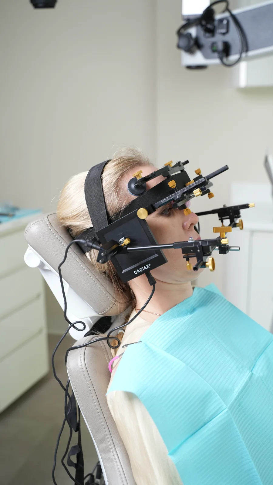
Imaging and movement recordings provide different information. Condylography records how your lower jaw moves as you follow a series of instructions. I use the Gamma CADIAX system for these recordings.
The traces and measurements help me examine jaw movement in detail. I also use them to set the articulator when planning a splint or dental restorations.
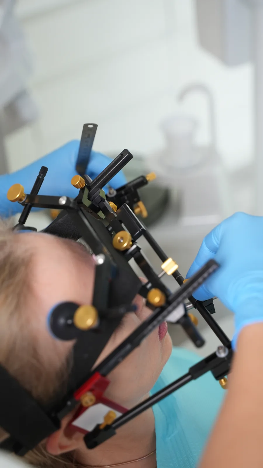
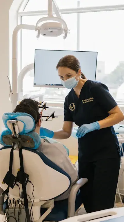
I fit and adjust the recording system, then ask you to open and close your mouth, move your lower jaw forward and move it to either side. I check the quality of the recordings before analysing the traces.
Condylography does not use X-rays. It records function rather than producing an image of the tissues inside the joint.
Including fitting and removal of the system, the assessment usually takes around 30–40 minutes. I check attachment stability and repeatability of the recordings to distinguish movement characteristics from recording interference.
Positioning and adjustment before recording.
After recording
How the findings are used
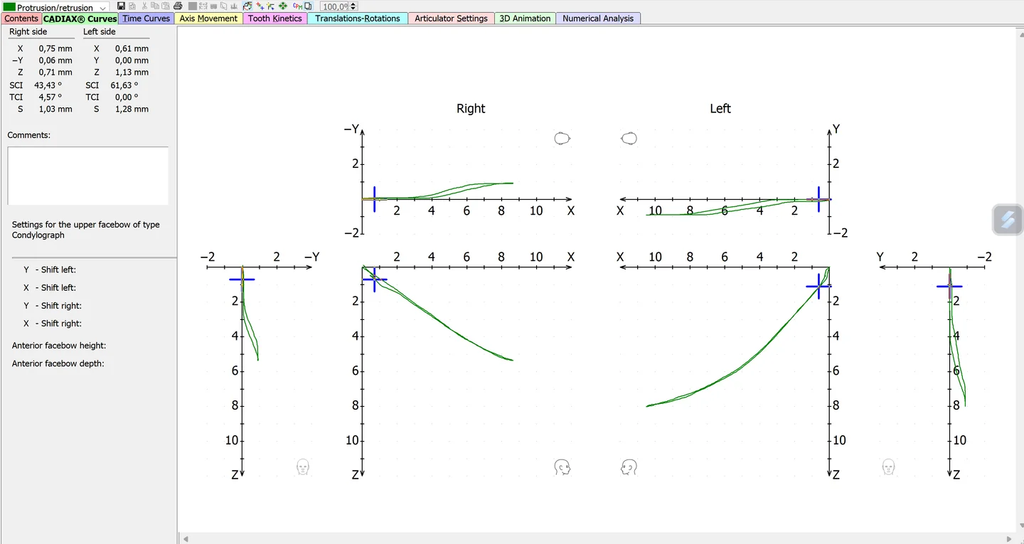
The example below shows protrusion of the lower jaw and its return. Right and left sides are displayed separately in several projections. I compare the paths, their direction and repeatability, and explain how these observations relate to the examination findings.
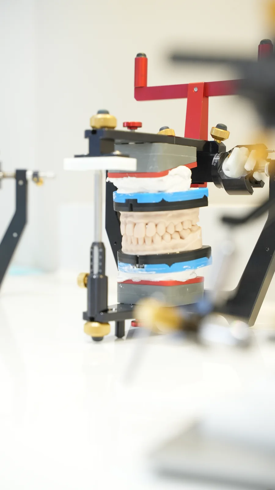
Individual parameters obtained during assessment are used to set the articulator. The dentist and technician work with jaw models in this device to plan the splint surface or the shape of future restorations. The recording is kept in the clinical record for comparison at later stages.
123
Jaw protrusion and return: a CADIAX recording from practice. Select the image to enlarge.
Right — recording from the patient’s right side.
Left — recording from the patient’s left side.
The X, Y and Z axes show different movement projections. Green traces record the outward and return paths.
Magnetic resonance imaging
How MRI helps assess the jaw joint
Magnetic resonance imaging (MRI) allows assessment of the joint disc and other soft tissues. It can show the position and shape of the disc, its relationship to the joint head and fluid within the joint.
CBCT helps assess bone, condylography records movement, and MRI adds information about internal soft tissues. I compare the images with your symptoms and examination findings to understand which findings matter for diagnosis and treatment planning.
In complex cases, I need MRI results to clarify the diagnosis and plan treatment. I also assess and treat patients who have contraindications to MRI or are not ready to undergo the examination for other reasons. In these situations, we discuss what other methods can tell us, which questions remain unanswered and how this affects treatment choices.
First, we establish the clinical problem: where pain occurs, what restricts movement and which findings need treatment. Depending on the examination, further records may include condylography, CBCT, MRI, photographs and digital models. We document your starting symptoms and function so that progress can be assessed later.
Stage 02
The treatment plan and its goals
I explain the findings and the proposed options. We identify what matters for monitoring: pain when chewing, mouth opening, locking episodes, tooth condition and comfort during everyday activities. We discuss the order of treatment, how long each stage is expected to take and the costs involved. Additional investigations and treatment are not automatically included in the basic consultation fee.
Stage 03
Splint therapy and conservative care
The splint is made from impressions or scans and a jaw-position record. I give the laboratory instructions for its design and contacts. Fabrication typically takes 7–10 days after the necessary records are collected; the fitting date is agreed with the laboratory schedule in mind.
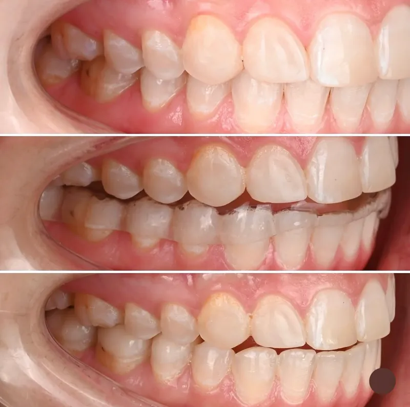
At the fitting appointment, I check that the appliance fits securely and that the opposing teeth meet and glide over its surface as intended. I show you how to insert and remove it, explain cleaning and prescribe a wear schedule. A repositioning splint helps maintain the lower jaw in a treatment position selected after assessment. I use a smooth-surface muscle-relaxation splint to help manage chewing muscle tension and more often make it for the lower jaw. The wear schedule depends on the design and treatment goal.
Reviews are commonly scheduled every 2–4 weeks. At each visit I assess pain, mouth opening, muscles and splint contacts. The findings determine whether to adjust the appliance, change the schedule or move to the next stage. When appropriate, I also recommend exercises and explain how to reduce strain on your jaw.
When to contact me before your next review. If the appliance causes pain, stop using it and contact me. A new change in how your teeth meet also needs review; do not adjust the appliance yourself.
Splint therapy: the teeth without a splint and with a lower-jaw splint. Photographs from one clinical case.
Stage 04
Dental rehabilitation
In my practice, treatment often includes dental rehabilitation to address damaged or missing teeth, extensive wear and problems with how the teeth meet. This may involve orthodontic treatment, prosthodontic treatment or a combination of both.
We first assess the space available for restoring tooth shape and the remaining tissue. If teeth need to move to make room for the planned restorations, we plan an orthodontic stage. Onlays, crowns and other restorations are designed using the functional records and agreed tooth positions.
A diagnostic wax-up models the proposed tooth shapes before the final restorations are made. For extensive treatment, temporary restorations let you try these shapes while eating and speaking. I use your feedback and the examination findings to refine the final design. The amount of restorative work depends on the condition of your teeth and what needs to be treated.
What to discuss before rehabilitation. Orthodontic treatment can cause tooth soreness and shortening of tooth roots; poor hygiene can lead to enamel damage and decay. Preparing a tooth for a crown is irreversible, and sensitivity or a need for root canal treatment may follow. Risks depend on the condition of your teeth and the proposed treatment. We discuss alternatives and the extent of treatment before starting.
Using dental models in functional treatment planning.
Stage 05
Review and ongoing monitoring
At follow-up appointments, I compare your symptoms and jaw movement with the records taken before treatment. I also ask how comfortable it is to eat, speak and open your mouth. Together, these observations help me decide whether to continue treatment, adjust it or move to regular check-ups.
Training & experience
My training and clinical work in TMJ care
Research into the functional diagnosis of TMJ disorders
2023 · PhD
Doctoral research on functional diagnosis
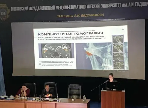
In 2023, I defended my dissertation on the choice of functional diagnostic methods for patients with TMJ disorders. The subject relates directly to a question that arises in clinical practice: which investigations are useful for this patient, and how should the findings inform treatment?
My co-authored research has focused on selecting methods to record jaw position and digitally fabricating individual diagnostic appliances. These records help us design diagnostic appliances and communicate the recorded jaw position to the dental laboratory.
Research publications ↗Training at the Vienna School of Interdisciplinary Dentistry (VieSID).
2023–2026 · VieSID
Training at VieSID in Vienna
My education includes the VieSID Basic Curriculum in 2023–2024 and Therapy Continuum in 2025–2026. Professor Rudolf Slavicek’s functional approach considers how the teeth, chewing muscles and jaw joints work together. It provides a framework for collecting clinical and instrumental findings and incorporating jaw function into dental treatment planning.
In an appointment, this means looking beyond the condition of an individual tooth. I also examine how your jaw moves, where pain occurs and which activities bring on discomfort.
I lead MedPalm Dental, the dental division of MedPalm Clinic in Jumeirah 1. When a plan involves several dental needs, I coordinate with colleagues in the relevant areas, including restorative dentistry, orthodontics, endodontics and surgical preparation where required. You should understand the sequence: what we are addressing now, who is responsible for the next stage and how progress will be reviewed.
Team lead
Dr. Svetlana Prisiazhnykh
Specialist Prosthodontist
11 years of experience
BDSMScPhDVieSID
PhD on functional diagnostics of TMJ disorders
1st place for TMD clinical work in 2025 and 2026 — Aesthetic Dentistry MENA Awards
Functional diagnostics using the VieSID approach and Gamma CADIAX
Yes. Conservative treatment is the main focus of my TMJ practice. The options depend on the diagnosis and care you have already received. I explain which approaches are suitable and when a surgeon’s involvement is needed.
Does everyone need condylography?
I start with a clinical examination. I recommend movement recordings when I need a closer look at jaw function or measurements to help plan treatment.
Is a night guard the same as a therapeutic splint?
Appliances may look similar but differ in their purpose, design and wear schedule. You should know what your appliance is intended to do, when to wear it and how the treatment will be monitored.
How long does treatment take?
If your plan includes splint therapy, fabrication usually takes 7–10 days after impressions or scans and a jaw-position record have been taken. Follow-up visits are usually scheduled every 2–4 weeks. I assess pain, mouth opening and splint contacts, adjusting the appliance and wear schedule as needed. We discuss the overall treatment duration after diagnosis; it depends on the joint and muscle findings, your response to care and whether further dental rehabilitation is needed.
Does clicking mean my joint is damaged?
Not necessarily. Clicking without pain or restricted movement usually does not need treatment. Pain, locking or difficulty opening your mouth should be assessed. The sound alone does not establish a diagnosis.
Can I have braces or crowns if my jaw hurts?
Before braces or crowns, I recommend finding the cause of your jaw pain and checking the joints and muscles. We can then agree with your orthodontist and prosthodontist which treatment should come first.
Can Botox replace assessment or splint therapy?
Botulinum toxin affects muscle activity. Any role for injections should be discussed after assessment; they do not replace evaluation of the joints, teeth and function. Evidence for TMD pain relief is mixed, so potential benefits, risks and alternatives need to be considered.
Existing scans and reports, a list of medications, your questions and any splint you currently use.
What we discuss
We discuss your symptoms, previous treatment and what I find during the examination. Once the assessment is complete, we talk through your treatment plan and the costs involved.
Where to find me
MedPalm Dental, Villa 6, 189B Jumeirah St, Jumeirah 1. Free private parking is available. Appointments are in English and Russian.
Villa 6, 189B Jumeirah St Jumeirah 1, Dubai, UAE Get directions ↗
Opening hours
Mon–Fri 9 am–8 pm; Sat–Sun 9 am–7 pm
Dr. Svetlana Explains
Bruxism and jaw pain need an individual assessment. Bite alignment alone does not establish the cause of pain; the effectiveness of botulinum toxin for TMD pain remains uncertain.